Liver fluke, transverse section

![]()
Wall Art and Photo Gifts from Science Photo Library
Liver fluke, transverse section
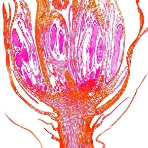
Liver fluke. Light micrograph of a transverse section through the anterior half of a liver fluke (Fasciola hepatica), a hermaphrodite tremotode worm and endoparasite that infects sheep. This section is in the region of the female reproductive organs. At far right, under the cuticle, are the vitaline glands (oval pink circles). Moving to the left, the dark purple-red circles are intestinal diverticula. Next are circular cavities (brown and black), which are the ovaries and eggs. The central area shows sections of the gut (purple). The large empty circles at left are parts of the uterus, some of which contain eggs. Magnification: x36 when printed at 10 centimetres across
Science Photo Library features Science and Medical images including photos and illustrations
Media ID 6469719
© DR KEITH WHEELER/SCIENCE PHOTO LIBRARY
Cross Section Diverticulum Eggs Fasciola Hepatica Flat Worm Fluke Histological Histology Intestinal Intestines Liver Fluke Ovaries Ovary Ovum Platyhelminthes Re Production Reproductive Transverse Uterus Light Micrograph Light Microscope Section Sectioned
EDITORS COMMENTS
This print showcases a transverse section of a liver fluke, specifically the anterior half of Fasciola hepatica. As an endoparasite that primarily infects sheep, this hermaphrodite trematode worm is both intriguing and formidable. The image focuses on the female reproductive organs within the worm. Starting from the far right under the cuticle, we can observe oval pink circles known as vitaline glands, which play a crucial role in sustaining this parasite's survival. Moving towards the left side of the image, one can notice dark purple-red circles representing intestinal diverticula. These structures aid in digestion and nutrient absorption for this fascinating creature. The circular cavities seen in brown and black hues are none other than ovaries filled with eggs - a pivotal part of its reproductive cycle. In addition to these ovaries, sections of the gut are visible in shades of purple at the central area. Towards the leftmost region lies large empty circles that belong to parts of its uterus; some even contain eggs waiting to be released into their host environment. This intricate cross-section provides valuable insights into not only liver flukes but also platyhelminthes or flatworms as a whole. With a magnification level equivalent to 36 times when printed at 10 centimeters across, this light micrograph offers an extraordinary view into nature's microscopic wonders while highlighting various aspects such as reproduction and anatomy within these mesmerizing creatures.
MADE IN THE USA
Safe Shipping with 30 Day Money Back Guarantee
FREE PERSONALISATION*
We are proud to offer a range of customisation features including Personalised Captions, Color Filters and Picture Zoom Tools
SECURE PAYMENTS
We happily accept a wide range of payment options so you can pay for the things you need in the way that is most convenient for you
* Options may vary by product and licensing agreement. Zoomed Pictures can be adjusted in the Cart.