Home > Science > SEM
Retina layers, SEM C018 / 0520

![]()
Wall Art and Photo Gifts from Science Photo Library
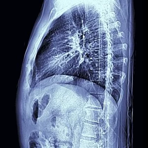
Retina layers, SEM C018 / 0520
Retina layers, coloured scanning electron micrograph (SEM). The retina of the human eye is found on the inside of the eyeball and is formed from a number of layers. The light-detecting part is formed of rod and cone photoreceptor cells. The lower third of this image contains the blood vessels and nerves that form the upper layer. The light entering our eyes has to penetrate though this layer to reach the tightly packed photosensitive outer segments of the photoreceptors (top third of image) which lie just below a layer of retinal pigment epithelium cells (top). Magnification: x480 when printed at 10 centimetres across
Science Photo Library features Science and Medical images including photos and illustrations
Media ID 9236877
© LOUISE HUGHES/SCIENCE PHOTO LIBRARY
Blood Vessels Colored Cone Cell Cones Epithelium Layer Layers Nerves Neural Ophthalmic Ophthalmology Photoreceptor Photoreceptors Physiological Physiology Retina Rod Cell Rods Sense Sensory Sight Tissue Vascular Vision Cells Nervous System Section Sectioned
FEATURES IN THESE COLLECTIONS
EDITORS COMMENTS
This print titled "Retina Layers" offers a mesmerizing glimpse into the intricate structure of the human eye. Captured using a scanning electron microscope (SEM), this colored image showcases the various layers that compose the retina, which lines the inner surface of our eyeballs. The lower portion of this image reveals a network of blood vessels and nerves, forming the upper layer of the retina. Light entering our eyes must traverse through this complex web to reach its destination: the densely packed outer segments of rod and cone photoreceptor cells, depicted in vibrant detail at the top third of this print. Just beneath these photosensitive cells lies another layer composed of retinal pigment epithelium cells. Together, these layers work harmoniously to facilitate light detection and transmit visual information to our brain for processing. With a magnification level equivalent to 480 times when printed at 10 centimeters across, this photograph provides an awe-inspiring view into one aspect of human anatomy rarely seen by naked eyes alone. It serves as a testament to both scientific advancement and artistic appreciation for nature's incredible design. Louise Hughes from Science Photo Library has skillfully captured not only an anatomical marvel but also an aesthetic masterpiece that invites contemplation on how we perceive and interpret our world through vision.
MADE IN THE USA
Safe Shipping with 30 Day Money Back Guarantee
FREE PERSONALISATION*
We are proud to offer a range of customisation features including Personalised Captions, Color Filters and Picture Zoom Tools
SECURE PAYMENTS
We happily accept a wide range of payment options so you can pay for the things you need in the way that is most convenient for you
* Options may vary by product and licensing agreement. Zoomed Pictures can be adjusted in the Cart.