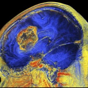
Brain MRI scan with Alzheimers QR code

![]()
Wall Art and Photo Gifts from Science Photo Library
Brain MRI scan with Alzheimers QR code
White matter fibres overlaid a mri scan of the human head. Coloured 3D diffusion spectral imaging (DSI) scan of the bundles of white matter nerve fibres in the brain. The fibres transmit nerve signals between brain regions and between the brain and the spinal cord. Diffusion spectrum imaging (DSI) is a variant of magnetic resonance imaging (MRI) in which a magnetic field maps the water contained in neuron fibers, thus mapping their criss-crossing patterns. A similar technique called diffusion tensor imaging (DTI) is also used to explore neural data of white matter fibres in the brain. Both methods allow mapping of their orientations and the connections between brain regions. Data/software: NIH Human Connectome Project /humanconnectomeproject.org)
Science Photo Library features Science and Medical images including photos and illustrations
Media ID 9224529
© PASIEKA/SCIENCE PHOTO LIBRARY
Brain Scan Connections Diffusion Spectral Imaging Diffusion Tensor Imaging Dsi Scan Dti Scan Fibers Fibre Tracking Fibres Human Brain Magnetic Resonance Imaging Mapping Mri Scan Nerve Bundles Nerve Fibre Pathway Pathways White Matter Nervous System
EDITORS COMMENTS
This print showcases the intricate complexity of the human brain through a combination of cutting-edge imaging techniques. The image features a Brain MRI scan with an Alzheimer's QR code, overlaid with colored 3D diffusion spectral imaging (DSI) scan of white matter nerve fibers. White matter fibres play a crucial role in transmitting nerve signals between different regions of the brain and connecting it to the spinal cord. By utilizing diffusion spectrum imaging (DSI), scientists can map these fibers' criss-crossing patterns by tracking water molecules within neuron fibers using magnetic resonance imaging (MRI). The technique used here is similar to diffusion tensor imaging (DTI), which also explores neural data related to white matter fibres in the brain. Both methods enable researchers to map fiber orientations and connections between various brain regions, providing valuable insights into how information is processed and transmitted throughout this remarkable organ. This awe-inspiring visual representation was made possible thanks to data and software from the NIH Human Connectome Project, a groundbreaking initiative aimed at mapping the structural and functional connectivity of the human brain. Through this mesmerizing artwork captured by PASIEKA/SCIENCE PHOTO LIBRARY, we are reminded of both the beauty and complexity that lies within our own minds.
MADE IN THE USA
Safe Shipping with 30 Day Money Back Guarantee
FREE PERSONALISATION*
We are proud to offer a range of customisation features including Personalised Captions, Color Filters and Picture Zoom Tools
SECURE PAYMENTS
We happily accept a wide range of payment options so you can pay for the things you need in the way that is most convenient for you
* Options may vary by product and licensing agreement. Zoomed Pictures can be adjusted in the Cart.