Home > Animals > Mammals > Muridae > Water Mouse
Corpus callosum, DTI MRI scan C017 / 7043

![]()
Wall Art and Photo Gifts from Science Photo Library
Corpus callosum, DTI MRI scan C017 / 7043
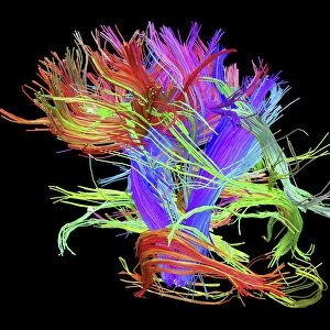
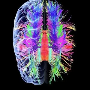
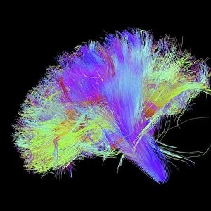
Corpus callosum. Coronal 3D diffusion tensor imaging (DTI) magnetic resonance imaging (MRI) scan of nerve pathways (red) in the brains corpus callosum. The corpus callosum is the band of nerve fibres that connects the two hemispheres of the brain (left and right). Diffusion tensor imaging measures the direction of water diffusion, which in the brain reveals the orientation of nerve fibres. The technique is also known as tractography, with the resulting image known as a tractogram
Science Photo Library features Science and Medical images including photos and illustrations
Media ID 9339881
© SHERBROOKE CONNECTIVITY IMAGING LAB/SCIENCE PHOTO LIBRARY
Brain Imaging Brain Scan Central Nervous System Cerebral Corpus Callosum Diffusion Tensor Imaging Dti Scan Fiber Fibers Fibre Fibres Imaging Technique Magnetic Resonance Imaging Mri Scan Mri Scanner Nerve Nerve Fibre Nerves Neural Pathway Neural Tract Paths Pathway Pathways Structural Tractogram Tractography White Matter Brain Neurological Neurology
FEATURES IN THESE COLLECTIONS
> Animals
> Mammals
> Muridae
> Water Mouse
EDITORS COMMENTS
This print showcases the intricate beauty of the human brain's corpus callosum, a vital structure that connects the left and right hemispheres. The image, captured using advanced diffusion tensor imaging (DTI) magnetic resonance imaging (MRI), reveals the complex network of nerve pathways within this crucial band of fibers. In this striking coronal 3D scan, the nerve pathways are highlighted in vibrant red against a deep black background. DTI measures water diffusion direction to unveil the orientation of these nerve fibers, providing valuable insights into brain connectivity. This technique, also known as tractography, produces a visually stunning tractogram that aids in understanding neural communication. The photograph not only emphasizes the structural elegance of our central nervous system but also underscores its significance for healthy biological functioning. It offers an exceptional glimpse into neurology and medicine by illustrating how different regions of our brain communicate through these neural tracts. As we explore this mesmerizing image further, it becomes evident that it represents more than just scientific research; it symbolizes humanity's relentless pursuit to unravel the mysteries hidden within ourselves. With each fiber meticulously mapped out by cutting-edge technology at Sherbrooke Connectivity Imaging Lab/Science Photo Library, this print serves as a testament to both human ingenuity and our unyielding curiosity about our own anatomy.
MADE IN THE USA
Safe Shipping with 30 Day Money Back Guarantee
FREE PERSONALISATION*
We are proud to offer a range of customisation features including Personalised Captions, Color Filters and Picture Zoom Tools
SECURE PAYMENTS
We happily accept a wide range of payment options so you can pay for the things you need in the way that is most convenient for you
* Options may vary by product and licensing agreement. Zoomed Pictures can be adjusted in the Cart.