Home > Science Photo Library > Human Body
Nerve cell growth

![]()
Wall Art and Photo Gifts from Science Photo Library
Nerve cell growth
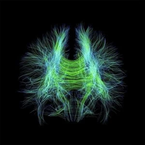
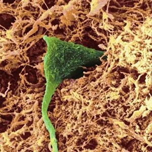
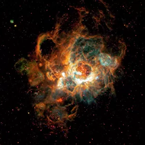
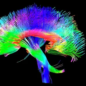
Nerve cell growth. Light micrograph of nerve cells (neurons) with immunofluorescent staining. These cells have been grown in culture. The stains show neurites (thin strands, either axons or dendrites) connecting nerve cell bodies. Short neurites are green, long ones are pink. Nerve cell bodies are blue, but non-neuronal cell nuclei (also blue) are seen as well. Neurons transmit electrical signals around the body, especially the brain and the spinal cord. These neurons have been grown from NT2 cells, a human teratocarcinoma cell line capable of differentiation into nerve cells. Such research may make neural regeneration possible for treating spinal paralysis and nerve disorders
Science Photo Library features Science and Medical images including photos and illustrations
Media ID 6421994
© FRANCOIS PAQUET-DURAND/SCIENCE PHOTO LIBRARY
Axon Axons Body C Ulture Cultured Dendrite Dendrites Immunofluorescence Nerve Nerve Cells Neurite Neurites Neuron Neurone Neurones Neurons Neuroscience Nucleus Stained Staining Light Micrograph Light Microscope Nervous System Neurology
EDITORS COMMENTS
This print showcases the intricate beauty of nerve cell growth. Through immunofluorescent staining, the image reveals a mesmerizing network of neurons that have been cultivated in culture. The vibrant colors highlight the neurites, which are thin strands connecting the nerve cell bodies. Short neurites glow with a vivid green hue, while longer ones emanate a delicate pink shade. The blue-colored nerve cell bodies stand out against their surroundings, and interestingly, non-neuronal cell nuclei can also be observed in blue. These remarkable cells play a crucial role in transmitting electrical signals throughout the body, particularly within the brain and spinal cord. What makes this image even more extraordinary is its connection to groundbreaking research. The neurons depicted here have been derived from NT2 cells—a human teratocarcinoma cell line capable of differentiating into nerve cells. This advancement offers hope for neural regeneration as a potential treatment for spinal paralysis and various nerve disorders. As we delve deeper into understanding the complexities of our nervous system, images like this one serve as visual reminders of both our progress and boundless possibilities within neuroscience. Science Photo Library has expertly captured this momentous snapshot that not only celebrates scientific exploration but also ignites curiosity about what lies ahead in unlocking the mysteries of our own biology.
MADE IN THE USA
Safe Shipping with 30 Day Money Back Guarantee
FREE PERSONALISATION*
We are proud to offer a range of customisation features including Personalised Captions, Color Filters and Picture Zoom Tools
SECURE PAYMENTS
We happily accept a wide range of payment options so you can pay for the things you need in the way that is most convenient for you
* Options may vary by product and licensing agreement. Zoomed Pictures can be adjusted in the Cart.