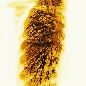
Heart and lungs, historical illustration

![]()
Wall Art and Photo Gifts from Science Photo Library
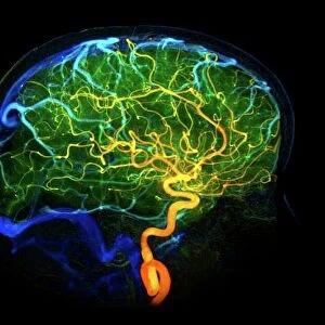
Heart and lungs, historical illustration
Heart and lungs, historical illustration. 19th Century hand coloured lithographic print showing the lungs (left and right) situated either side of the heart (lower centre). The pulmonary veins (red) and arteries (blue) can be seen leading to and from the heart respectively. The trachea (wind pipe, white) can be seen running down from the larynx (voicebox, top centre) in the neck, which branches into the two bronchi and then further into numerous bronchioles inside the lungs. Image from Traite complet de l anatomie de l homme, comprenant la medecine operatoire Vol. 4 (1836), by Jean-Baptiste Marc Bourgery and illustrated by Nicolas-Henri Jacob
Science Photo Library features Science and Medical images including photos and illustrations
Media ID 6327959
© SCIENCE PHOTO LIBRARY
1836 Airway Airways Alveolus Aorta Arteries Back Bronchi Bronchiole Bronchioles Bronchus Chest Circulatory Descriptive Anatomy Diagram French Interior Internal Larynx Lithograph Lithographic Print Lung Lungs Neck Organs Pulmonary Pulmonary System Respiratory System Thoracic Thorax Throat Trachea Vascular Veins Vessels Vol 4 Volume Four Artery Cardiovascular System Circulation Plate 5 Vein
EDITORS COMMENTS
This 19th-century hand-coloured lithographic print showcases the intricate beauty of the human heart and lungs. The image, taken from Jean-Baptiste Marc Bourgery's "Traite complet de l'anatomie de l'homme" offers a detailed view of the chest's interior, revealing the complex network of veins, arteries, and organs that make up our respiratory and circulatory systems. In this remarkable illustration, we observe the lungs gracefully positioned on either side of the heart at its lower center. The vibrant red pulmonary veins elegantly transport oxygen-rich blood back to the heart, while blue arteries carry oxygen-depleted blood away from it. Above them lies the trachea or windpipe in white, extending down from the voicebox in the neck. The artist's meticulous attention to detail is evident as they depict how these airways branch into bronchi and further divide into countless bronchioles within each lung. This comprehensive representation provides a visual understanding of how our respiratory system functions. With its historical significance and artistic flair, this lithograph not only serves as an educational tool for studying anatomy but also stands as a testament to scientific exploration during that era. It reminds us of our body's incredible complexity while highlighting both its fragility and resilience. Science Photo Library presents this stunning piece that combines artistry with scientific accuracy—a true treasure for those fascinated by biology, history, or simply captivated by humanity's internal wonders.
MADE IN THE USA
Safe Shipping with 30 Day Money Back Guarantee
FREE PERSONALISATION*
We are proud to offer a range of customisation features including Personalised Captions, Color Filters and Picture Zoom Tools
SECURE PAYMENTS
We happily accept a wide range of payment options so you can pay for the things you need in the way that is most convenient for you
* Options may vary by product and licensing agreement. Zoomed Pictures can be adjusted in the Cart.