Home > Europe > United Kingdom > Scotland > Moray > Keith
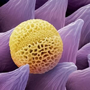
Water lily leaf, light micrograph

![]()
Wall Art and Photo Gifts from Science Photo Library
Water lily leaf, light micrograph
Water lily leaf. Light micrograph of a transverse section through the leaf of a water lily (Nympha sp.) plant. All aquatic plants (hydrophytes) have a similar structure. The upper epidermis of the leaf has a thin cuticle (top) underneath which is a multi-layered palisade mesophyll. In-between the palisade cells are elongated sclereids (purple) for support. Underneath this is the spongy mesophyll and large intercellular air spaces (lacunae, white). The vascular bundles (dense patches) consist of xylem (red) and phloem (dark-blue ovals). The base of the midrib, under the epidermis, consists of collenchyma cells (dark blue). Magnification: x103 when printed 10 centimetres wide
Science Photo Library features Science and Medical images including photos and illustrations
Media ID 6282014
© DR KEITH WHEELER/SCIENCE PHOTO LIBRARY
Bundle Bundles Cell Biology Collenchyma Cuticle Cytology Hydrophyte Hydrophytic Intercellular Lacuna Lacunae Mid Rib Palisade Mesophyll Permeable Phloem Plant Structure Spaces Spongy Mesophyll Support Supportive Vascular Bundle Vascular Tissue Xylem Cells Light Micrograph Light Microscope Section Sectioned
FEATURES IN THESE COLLECTIONS
> Animals
> Mammals
> Muridae
> Water Mouse
> Arts
> Realistic drawings
> Nature art
> Botanical artwork
> Europe
> United Kingdom
> Scotland
> Moray
> Keith
EDITORS COMMENTS
This print showcases the intricate beauty of a water lily leaf, captured through a light micrograph. The transverse section reveals the fascinating structure that is common among all aquatic plants. The delicate upper epidermis of the leaf is protected by a thin cuticle, while just beneath it lies a multi-layered palisade mesophyll. Within this layer, elongated sclereids can be seen in purple, providing essential support to the leaf. Moving further down, we encounter the spongy mesophyll and large intercellular air spaces known as lacunae in white. These spaces allow for efficient gas exchange within the leaf. The dense patches throughout represent vascular bundles consisting of xylem in red and phloem depicted as dark-blue ovals. These bundles play a crucial role in transporting water and nutrients throughout the plant. Examining closer at the base of the midrib under the epidermis, we observe collenchyma cells shown in dark blue which contribute to structural support. This stunning image magnified 103 times when printed 10 centimeters wide truly highlights nature's remarkable design. With its vibrant colors and intricate details, this print serves as a testament to both botanical wonders and scientific exploration. It invites us into an enchanting world where cell biology meets artistry—a visual celebration of hydrophytes' unique adaptations that thrive underwater.
MADE IN THE USA
Safe Shipping with 30 Day Money Back Guarantee
FREE PERSONALISATION*
We are proud to offer a range of customisation features including Personalised Captions, Color Filters and Picture Zoom Tools
SECURE PAYMENTS
We happily accept a wide range of payment options so you can pay for the things you need in the way that is most convenient for you
* Options may vary by product and licensing agreement. Zoomed Pictures can be adjusted in the Cart.