Home > Arts > Minimalist artwork > Monochrome artwork > Fine art
Heart and lung disease, X-ray C018 / 0498

![]()
Wall Art and Photo Gifts from Science Photo Library
Heart and lung disease, X-ray C018 / 0498
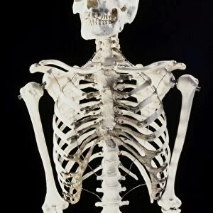
Heart and lung disease. Coloured X-ray of the chest of an 80 year old patient with heart and lung disease. The patient is a former miner and has chronic obstructive pulmonary disease (COPD) and an enlarged left ventricle (at centre right) of the heart. COPD is a collection of diseases that includes emphysema and chronic bronchitis. Damage to the lung tissue (seen as opaque plaques) reduces gas exchange in the lungs. The heart has to pump harder to get oxygen to the body. As it is a muscle, the heart grows if it has an increased work load. A pacemaker (upper right) has also been fitted, to supply electrical impulses to the heart to keep it beating regularly
Science Photo Library features Science and Medical images including photos and illustrations
Media ID 9237359
© ZEPHYR/SCIENCE PHOTO LIBRARY
Bronchitis Cardiac Cardiology Cardiomegaly Chest Device Diagnosis Diagnostic Eighties Elderly Electrical Electronic Emphysema Enlarged Enlargement Front Frontal Heart Disease Hypertrophic Implant Left Ventricle Lung Disease Lungs Miner Pacemaker Prosthesis Radiography Respiratory X Ray Machine Xray Abnormal Condition Disorder Eighty Occupational Disease Unhealthy
FEATURES IN THESE COLLECTIONS
> Arts
> Minimalist artwork
> Monochrome artwork
> Fine art
> Arts
> Minimalist artwork
> Monochrome artwork
> Monochrome paintings
EDITORS COMMENTS
This print showcases the intricate details of heart and lung disease in an 80-year-old patient. The subject, a former miner, is depicted with chronic obstructive pulmonary disease (COPD) and an enlarged left ventricle at the center right of the heart. COPD encompasses various conditions such as emphysema and chronic bronchitis, which have caused damage to the lung tissue visible as opaque plaques on the X-ray. The consequences of this condition are evident: reduced gas exchange in the lungs necessitates increased effort from the heart to supply oxygen to the body. As a result, we witness how this vital muscle has grown due to its heightened workload. To regulate its rhythm, a pacemaker has been implanted in the upper right portion of the chest, providing electrical impulses that ensure regular beating. This monochrome image not only serves as a diagnostic tool but also highlights both medical advancements and occupational hazards faced by individuals like miners throughout their lives. It underscores how long-term exposure to harmful substances can lead to severe health issues later in life. Through this photograph captured by ZEPHYR/SCIENCE PHOTO LIBRARY, we gain insight into complex cardiac and respiratory disorders while appreciating scientific progress aimed at improving patients' quality of life.
MADE IN THE USA
Safe Shipping with 30 Day Money Back Guarantee
FREE PERSONALISATION*
We are proud to offer a range of customisation features including Personalised Captions, Color Filters and Picture Zoom Tools
FREE COLORIZATION SERVICE
You can choose advanced AI Colorization for this picture at no extra charge!
SECURE PAYMENTS
We happily accept a wide range of payment options so you can pay for the things you need in the way that is most convenient for you
* Options may vary by product and licensing agreement. Zoomed Pictures can be adjusted in the Cart.